Bronchial Pattern Dog
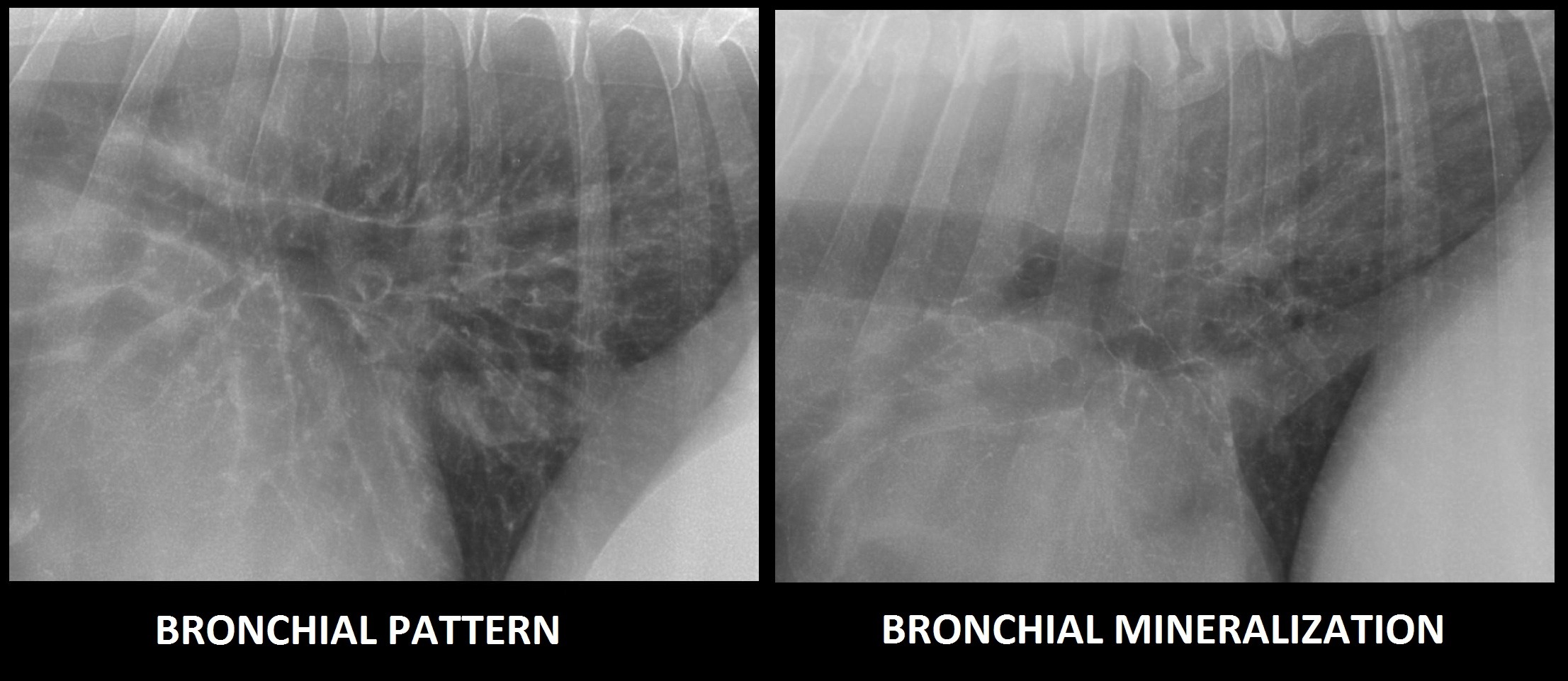
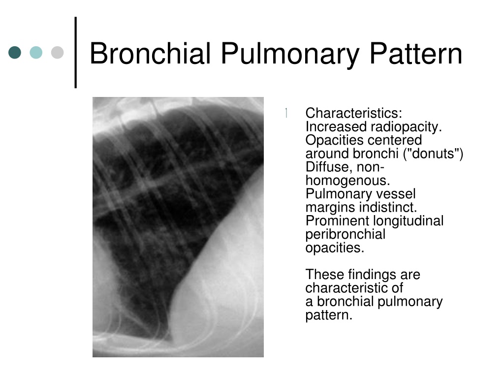
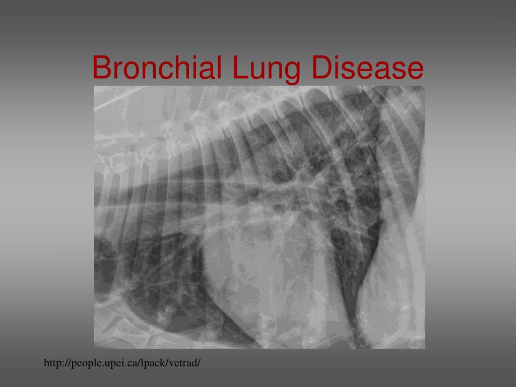
Bronchial Pattern Dog - Web b) bronchial patterns: Web when a dog breathes air in through its nose or mouth, the air travels down the trachea, which divides into the tubes known as the right and left bronchi, then into the smaller airways called bronchioles in the lungs. Perihilar distribution (in dogs) is most associated with congestive heart failure. Bacterial > allergic (eosinophilic) cats: It is discussed in this chapter as part of tracheobronchitis. Web tracheobronchitis is a sudden or longterm inflammation of the trachea and bronchial airways; Web alveolar patterns are typically fluffy and indistinct, and coalesce. If the cough lasts more than two months, it's generally referred to as chronic bronchitis. Matthew winter, dacvr will review the radiographic features of lung patterns in dogs and cats as well as the keys to interpreting the meaning of these patterns. It may also extend into the lungs. This pattern comes closest to helping shed light on what disease the pet is suffering from. This makes them easier to see, especially in the periphery of the lung (image 2). The thickening of those structures results in enhanced radiographic visualization of. Also see professional content regarding tracheobronchitis. Web alveolar patterns are typically fluffy and indistinct, and coalesce. Yellow circles) and parallel lines (“tramlines”; The walls are thickened due to a combination of smooth muscle hypertrophy, mucus production, cellular infiltrate, and in come cases (feline asthma), bronchoconstriction. Web tracheobronchitis is a sudden or longterm inflammation of the trachea and bronchial airways; It often occurs in dogs already affected by respiratory disease or a disorder of the lungs or airways. Diffuse interstitial or alveolar patters may be due to vasculitis, acute lung injury (ali), acute respiratory distress syndrome (ards), atypical pneumonia, or neoplasia such as lymphoma. Web when a dog breathes air in through its nose or mouth, the air travels down the trachea, which divides into the tubes known as the right and left bronchi, then into the smaller airways called bronchioles in the lungs. It may also extend into the lungs. To understand the disease, it's first important to know about the basic anatomy. It is discussed in this chapter as part of tracheobronchitis. Web b) bronchial patterns: Web a bronchial pattern on radiographs indicates a condition that involves the airways. The thickening of those structures results in enhanced radiographic visualization of. Web diffuse pulmonary disease may be in the form of a bronchial pattern, or interstitial or alveolar pattern. It often occurs in dogs already affected by respiratory disease or a disorder of the lungs or airways. It may also extend into the lungs. It can be a subtle pattern to recognize, so lets look at some of the features. Matthew winter, dacvr will review the radiographic features of lung patterns in dogs and cats as well as the. Diffuse interstitial or alveolar patters may be due to vasculitis, acute lung injury (ali), acute respiratory distress syndrome (ards), atypical pneumonia, or neoplasia such as lymphoma. This does not hold true in the cat. If the cough lasts more than two months, it's generally referred to as chronic bronchitis. Also see professional content regarding tracheobronchitis. Perihilar distribution (in dogs) is. Web when a dog breathes in, air flows through their mouth or nose to their trachea, also known as the windpipe. This makes them easier to see, especially in the periphery of the lung (image 2). In a true bronchial pattern that stems from infectious/inflammatory disease, the bronchial walls are thickened because of inflammatory tissue and cells surrounding the airways.. The walls are thickened due to a combination of smooth muscle hypertrophy, mucus production, cellular infiltrate, and in come cases (feline asthma), bronchoconstriction. Web diffuse pulmonary disease may be in the form of a bronchial pattern, or interstitial or alveolar pattern. It may also extend into the lungs. Typically, neither the esophagus nor tracheobronchial lymph nodes are visualized in thoracic. In a true bronchial pattern that stems from infectious/inflammatory disease, the bronchial walls are thickened because of inflammatory tissue and cells surrounding the airways. Web when a dog breathes in, air flows through their mouth or nose to their trachea, also known as the windpipe. This makes them easier to see, especially in the periphery of the lung (image 2).. The trachea then carries the inhaled air to the bronchi (the tubes that connect the. This does not hold true in the cat. The thickening of those structures results in enhanced radiographic visualization of. Web bronchitis in dogs is a common illness that affects the upper airways and causes coughing. Typically, neither the esophagus nor tracheobronchial lymph nodes are visualized. Web tracheobronchitis is a sudden or longterm inflammation of the trachea and bronchial airways; This pattern comes closest to helping shed light on what disease the pet is suffering from. Web bronchial lung pattern the bronchial pattern is obtained when the bronchial wall is infiltrated by cells or fluid or when the peribronchial space is replaced by cells or fluid.. It often occurs in dogs already affected by respiratory disease or a disorder of the lungs or airways. Web when a dog breathes in, air flows through their mouth or nose to their trachea, also known as the windpipe. Web bronchitis in dogs is a common illness that affects the upper airways and causes coughing. Web when a dog breathes. Web when a dog breathes air in through its nose or mouth, the air travels down the trachea, which divides into the tubes known as the right and left bronchi, then into the smaller airways called bronchioles in the lungs. It is discussed in this chapter as part of tracheobronchitis. Web bronchial patterns are generally distinct from interstitial and alveolar patterns, with the primary cause being thickening of the larger, conducting airways. This makes them easier to see, especially in the periphery of the lung (image 2). The thickening of those structures results in enhanced radiographic visualization of. Web when a dog breathes in, air flows through their mouth or nose to their trachea, also known as the windpipe. What are the signs of chronic bronchitis? Yellow circles) and parallel lines (“tramlines”; Bronchial pattern is caused by thickening and increased prominence of the bronchial walls, usually secondary to chronic inflammation. A bronchial pattern is an abnormal lung opacity caused by peribronchial cellular, fluid and fibrotic infiltration, or bronchial mucosal and submucosal thickening (chronic bronchitis). Web alveolar patterns are typically fluffy and indistinct, and coalesce. This pattern comes closest to helping shed light on what disease the pet is suffering from. Web diffuse pulmonary disease may be in the form of a bronchial pattern, or interstitial or alveolar pattern. This does not hold true in the cat. It often occurs in dogs already affected by respiratory disease or a disorder of the lungs or airways. Web bronchitis in dogs is a common illness that affects the upper airways and causes coughing.
Chronic & Persistent Coughing in a Dog Clinician's Brief

Radiographic Approach to the Coughing Pet • MSPCAAngell

Topographical distribution and radiographic pattern of lung lesions in
Radiographic Approach to the Coughing Pet • MSPCAAngell
PPT Veterinary Radiology PowerPoint Presentation, free download ID

Imaging the Coughing Dog
PPT Thoracic Radiology of the Dog PowerPoint Presentation, free

Thoracic radiograph of dog showed mild bronchial pattern (A) and an
Interpreting thoracic radiograph lung patterns VETgirl Veterinary

Topographical distribution and radiographic pattern of lung lesions in
Dogs And Cats With Respiratory Tract Disorders Can Present To Veterinarians For A Variety Of Clinical Signs Including Nasal Discharge, Sneeze, Reverse Sneeze, Noisy Breathing (Snoring/Stertor, Stridor, Wheeze), Cough, Alterations In Respiratory Rate Or Effort, And Respiratory Distress.
Cranioventral Distribution Is Most Associated With Bronchopneumonia;
If The Cough Lasts More Than Two Months, It's Generally Referred To As Chronic Bronchitis.
The Walls Are Thickened Due To A Combination Of Smooth Muscle Hypertrophy, Mucus Production, Cellular Infiltrate, And In Come Cases (Feline Asthma), Bronchoconstriction.
Related Post: