Pulmonary Disease Pattern Ecg
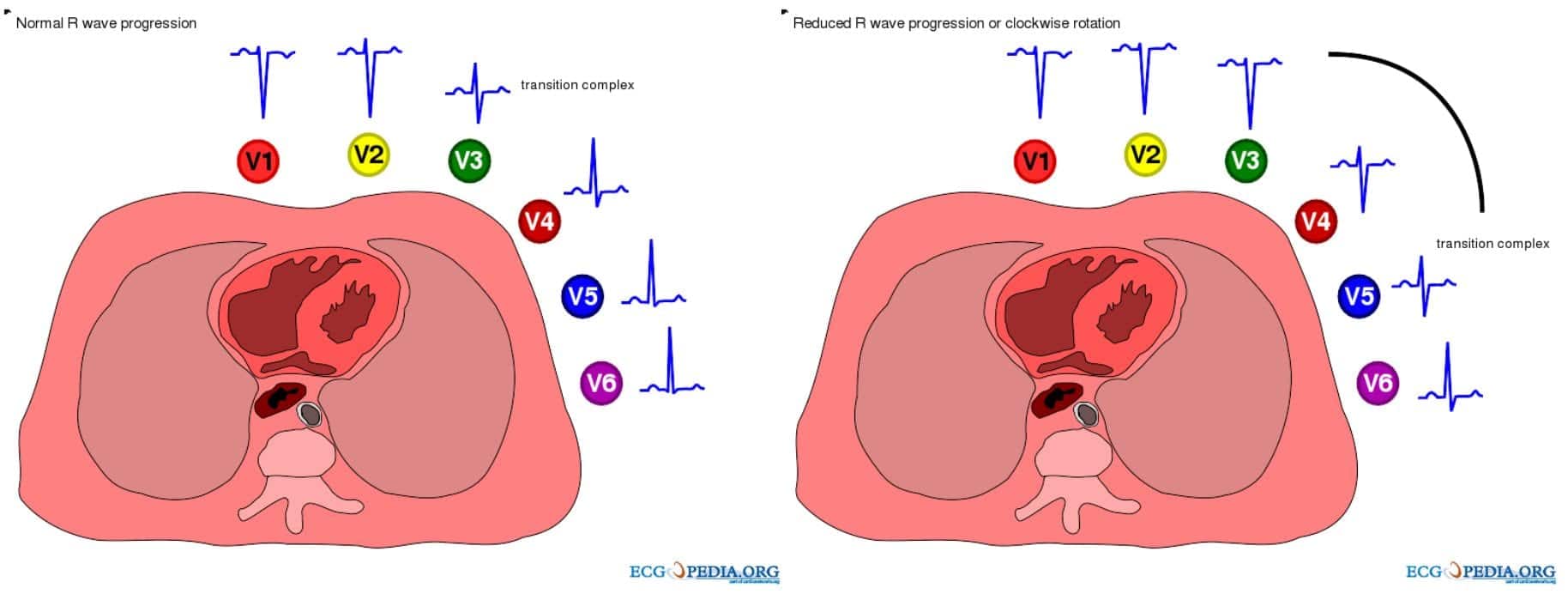
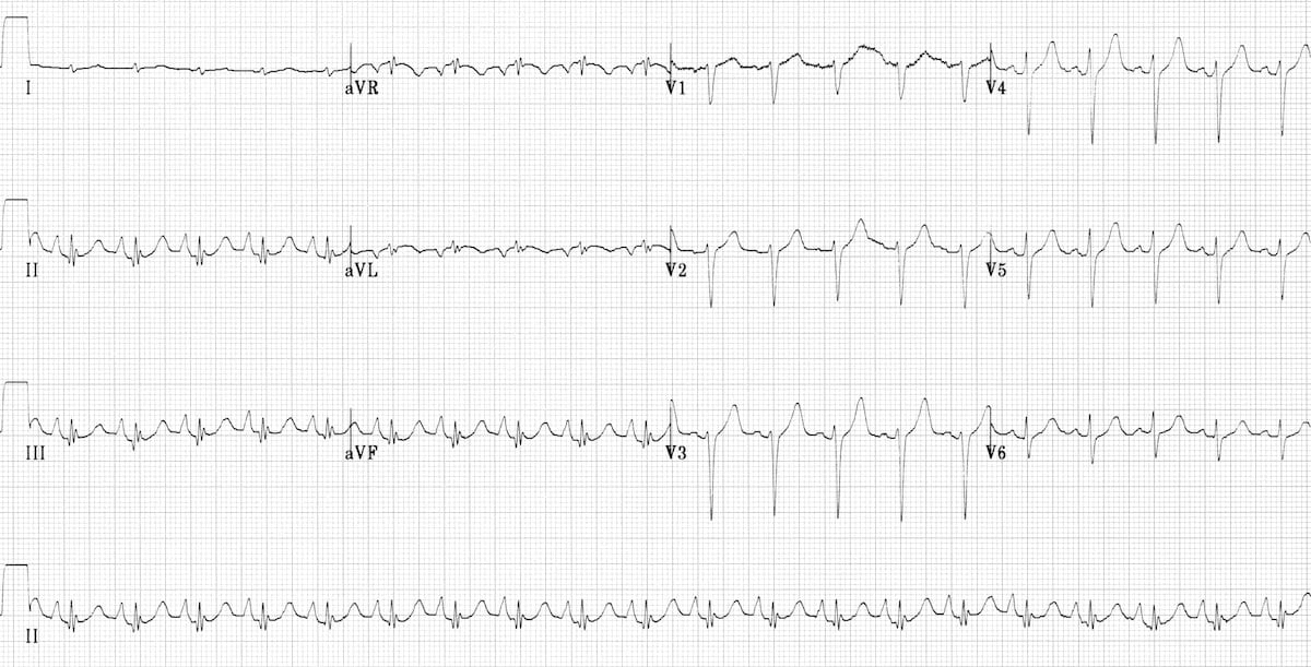
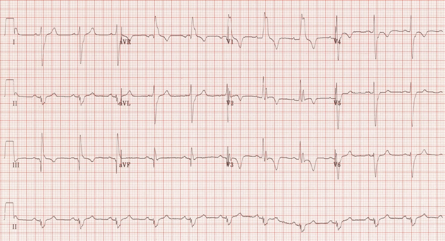
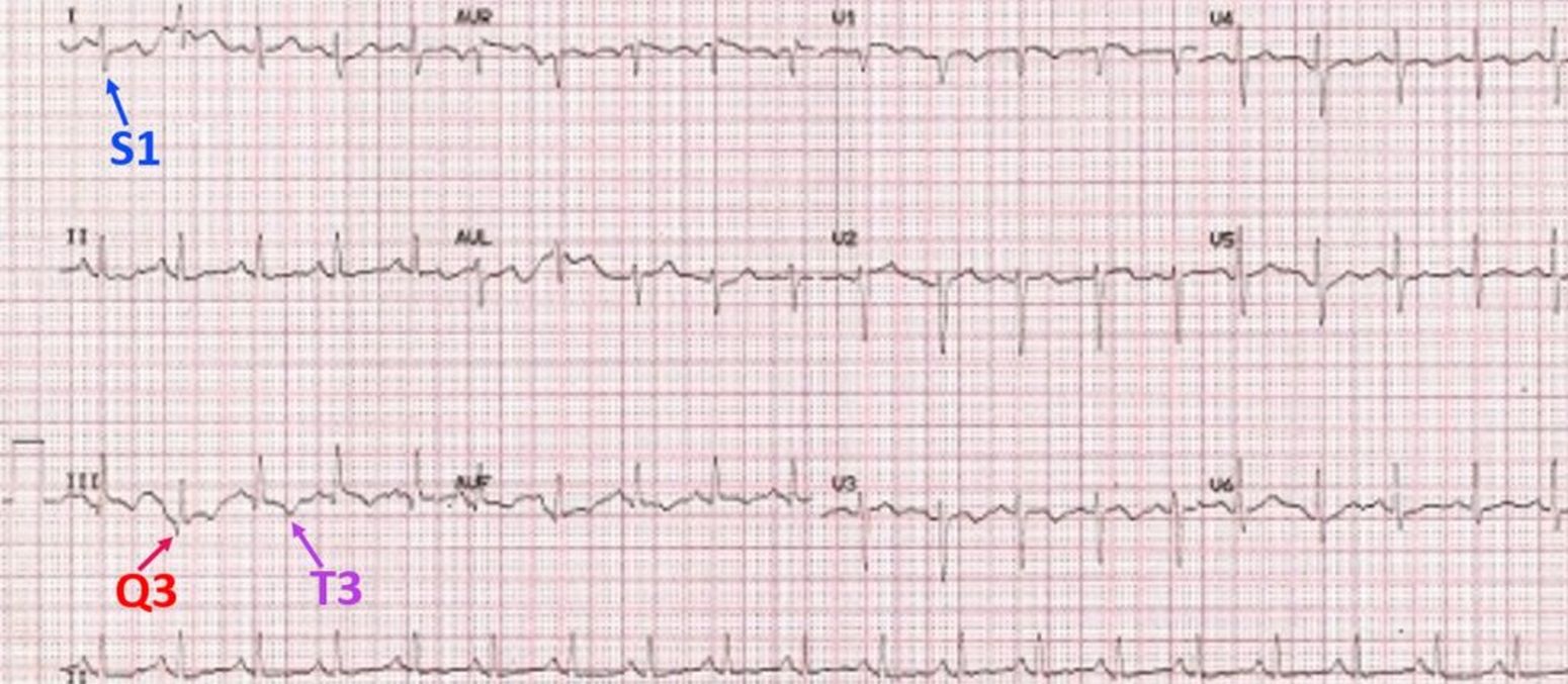
Pulmonary Disease Pattern Ecg - Web ecg abnormalities are common in patients with pulmonary embolism, with the most frequent being sinus tachycardia, right ventricular strain, and the classic s1q3t3 pattern. Web formal pulmonary function tests: Web this article has three purposes: Dilation of the right atrium and right ventricle with consequent shift in the position of the heart. Web ecg changes occur in chronic obstructive pulmonary disease (copd) due to: Web cvd indicates cardiovascular disease; Web electrocardiography (ecg) is a useful adjunct to other pulmonary tests because it provides information about the right side of the heart and therefore pulmonary disorders such as chronic pulmonary hypertension and pulmonary embolism. • right axis deviation or vertical axis of the qrs complex. First, to review the ecg patterns that suggest the presence of underlying copd; Web the electrocardiogram is often abnormal in patients who have chronic obstructive pulmonary disease. Ecg findings often suggest right ventricular pressure overload or strain. The presence of hyperexpanded emphysematous lungs within the chest; (2) an s 1 s 2 s 3 pattern, a relatively uncommon finding not highly specific for copd 13 that reflects an anomalous wave front rightward and superiorly. Our aim was to separate the effects on ecg by airway obstruction, emphysema and right ventricular (rv) afterload in patients with copd. To evaluate the extent and diagnostic values of ecg changes among copd patients suffering from broad spectrum of respiratory diseases. Web chronic obstructive pulmonary disease often causes a characteristic electrocardiographic pattern that reflects mainly the low diaphragm resulting from the increased lung volume. • right axis deviation of the p waves. Web this article has three purposes: • right axis deviation or vertical axis of the qrs complex. Web the electrocardiogram is often abnormal in patients who have chronic obstructive pulmonary disease. The presence of hyperexpanded emphysematous lungs within the chest; Web ecg abnormalities are common in patients with pulmonary embolism, with the most frequent being sinus tachycardia, right ventricular strain, and the classic s1q3t3 pattern. Ecg changes commonly associated with pulmonary diseases such as copd. Web cvd indicates cardiovascular disease; Web chronic obstructive pulmonary disease often causes a characteristic electrocardiographic pattern. Web chronic obstructive pulmonary disease often causes a characteristic electrocardiographic pattern that reflects mainly the low diaphragm resulting from the increased lung volume. Web formal pulmonary function tests: Web this article has three purposes: To evaluate the extent and diagnostic values of ecg changes among copd patients suffering from broad spectrum of respiratory diseases. Our aim was to separate the. • right axis deviation of the p waves. Web cvd indicates cardiovascular disease; Web in copd, the various pathophysiological mechanisms modify the ecg differently. Web electrocardiography (ecg) is a useful adjunct to other pulmonary tests because it provides information about the right side of the heart and therefore pulmonary disorders such as chronic pulmonary hypertension and pulmonary embolism. The prevalence. Web ecg changes occur in chronic obstructive pulmonary disease (copd) due to: Our aim was to separate the effects on ecg by airway obstruction, emphysema and right ventricular (rv) afterload in patients with copd. Web electrocardiography (ecg) is a useful adjunct to other pulmonary tests because it provides information about the right side of the heart and therefore pulmonary disorders. Web chronic obstructive pulmonary disease often causes a characteristic electrocardiographic pattern that reflects mainly the low diaphragm resulting from the increased lung volume. Dilation of the right atrium and right ventricle with consequent shift in the position of the heart. • right axis deviation of the p waves. Web electrocardiographic (ecg) findings may help in clinical decision making regarding this. Increased stimulation of the sympathetic nervous system due to pain, anxiety and hypoxia. Copd is associated with increased airway resistance, alveolar and pulmonary capillary destruction, air trapping, chronic hypoxemia and increased work of breathing. Our aim was to separate the effects on ecg by airway obstruction, emphysema and right ventricular (rv) afterload in patients with copd. Dilation of the right. Web electrocardiographic (ecg) findings may help in clinical decision making regarding this disease entity. Second,to describe the special ecg abnormalities that suggest acute superimposed illness (these have prognostic or therapeutic significance); • right axis deviation or vertical axis of the qrs complex. Web the following ecg signs reflecting ccp were collected: Web electrocardiography (ecg) is a useful adjunct to other. This pattern is characterized by a large s wave in lead i, a q wave in lead iii, and an inverted t wave in lead iii. Web electrocardiography (ecg) is a useful adjunct to other pulmonary tests because it provides information about the right side of the heart and therefore pulmonary disorders such as chronic pulmonary hypertension and pulmonary embolism.. Web cvd indicates cardiovascular disease; Web formal pulmonary function tests: Web based on the low voltage in leads v 1, v 2, v 3, the rightward frontal plane axis, incomplete right bundle branch block (rbbb), and persistent precordial s waves, the computer interpreted the overall pattern as consistent with pulmonary disease. The presence of hyperexpanded emphysematous lungs within the chest;. Web electrocardiographic (ecg) findings may help in clinical decision making regarding this disease entity. Web this article has three purposes: We found a similar pattern in the low. First, to review the ecg patterns that suggest the presence of underlying copd; Increased stimulation of the sympathetic nervous system due to pain, anxiety and hypoxia. Web based on the low voltage in leads v 1, v 2, v 3, the rightward frontal plane axis, incomplete right bundle branch block (rbbb), and persistent precordial s waves, the computer interpreted the overall pattern as consistent with pulmonary disease. This pattern is characterized by a large s wave in lead i, a q wave in lead iii, and an inverted t wave in lead iii. Web the electrocardiogram is often abnormal in patients who have chronic obstructive pulmonary disease. Screening for colorectal cancer screening for hypertension screening for lung cancer screening for prediabetes and type 2. Copd is associated with increased airway resistance, alveolar and pulmonary capillary destruction, air trapping, chronic hypoxemia and increased work of breathing. Our aim was to separate the effects on ecg by airway obstruction, emphysema and right ventricular (rv) afterload in patients with copd. • right axis deviation of the p waves. Web electrocardiography (ecg) is a useful adjunct to other pulmonary tests because it provides information about the right side of the heart and therefore pulmonary disorders such as chronic pulmonary hypertension and pulmonary embolism. (2) an s 1 s 2 s 3 pattern, a relatively uncommon finding not highly specific for copd 13 that reflects an anomalous wave front rightward and superiorly. The presence of hyperexpanded emphysematous lungs within the chest; Dilation of the right atrium and right ventricle with consequent shift in the position of the heart. Web the following ecg signs reflecting ccp were collected: Ecg changes commonly associated with pulmonary diseases such as copd. Ecgs were interpreted blindly in 63 patients with severe copd (group 1) versus 83 patients with mild or moderate copd (group 2). Ecg findings often suggest right ventricular pressure overload or strain. Web ecg changes in pe are related to:
Pulmonary Embolism (PE) Causes, symptoms, diagnosis, treatment
ECG in Chronic Obstructive Pulmonary Disease • LITFL • ECG Library
ECG in Chronic Obstructive Pulmonary Disease • LITFL • ECG Library
Pulmonary Embolism ECG New Health Advisor

Pulmonary Disease Pattern On Ekg Pattern.rjuuc.edu.np

The ECG's of Pulmonary Embolism Resus

Pulmonary embolism and S1Q3 pattern Cardiocases

S1Q3T3 on ECG in a patient with Acute Pulmonary Embolism GrepMed
pulmonary disease pattern ecg Hình ảnh có liên quan Diseases Club

ECG in Chronic Obstructive Pulmonary Disease • LITFL • ECG Library
To Evaluate The Extent And Diagnostic Values Of Ecg Changes Among Copd Patients Suffering From Broad Spectrum Of Respiratory Diseases.
Increased Stimulation Of The Sympathetic Nervous System Due To Pain, Anxiety And Hypoxia.
• Right Axis Deviation Or Vertical Axis Of The Qrs Complex.
Web This Article Has Three Purposes:
Related Post: