Pulmonary Disease Pattern Ekg
Pulmonary Disease Pattern Ekg - (see also electrocardiography in cardiovascular disorders.) Web around 18% of patients with pe will have a completely normal ecg. The presence of hyperexpanded emphysematous lungs within the chest; • right axis deviation or vertical axis of the qrs complex. Web in copd, the various pathophysiological mechanisms modify the ecg differently. • right axis deviation of the p waves. Our aim was to separate the effects on ecg by airway obstruction, emphysema and right ventricular (rv) afterload in patients with copd. Ecg findings often suggest right ventricular pressure overload or strain. Ecg changes commonly associated with pulmonary diseases such as copd. Web electrocardiography (ecg) is a useful adjunct to other pulmonary tests because it provides information about the right side of the heart and therefore pulmonary disorders such as chronic pulmonary hypertension and pulmonary embolism. The presence of hyperexpanded emphysematous lungs within the chest; Ecg findings often suggest right ventricular pressure overload or strain. Ecg changes commonly associated with pulmonary diseases such as copd. Our aim was to separate the effects on ecg by airway obstruction, emphysema and right ventricular (rv) afterload in patients with copd. Web ecg changes occur in chronic obstructive pulmonary disease (copd) due to: Web in copd, the various pathophysiological mechanisms modify the ecg differently. • right axis deviation or vertical axis of the qrs complex. Web around 18% of patients with pe will have a completely normal ecg. Web objective patients with chronic obstructive pulmonary disease (copd) often have abnormal ecgs. (see also electrocardiography in cardiovascular disorders.) (see also electrocardiography in cardiovascular disorders.) Ecg findings often suggest right ventricular pressure overload or strain. Web ecg changes occur in chronic obstructive pulmonary disease (copd) due to: The presence of hyperexpanded emphysematous lungs within the chest; Our aim was to separate the effects on ecg by airway obstruction, emphysema and right ventricular (rv) afterload in patients with copd. Ecg changes commonly associated with pulmonary diseases such as copd. Our aim was to separate the effects on ecg by airway obstruction, emphysema and right ventricular (rv) afterload in patients with copd. Web electrocardiography (ecg) is a useful adjunct to other pulmonary tests because it provides information about the right side of the heart and therefore pulmonary disorders such as. • right axis deviation of the p waves. Web objective patients with chronic obstructive pulmonary disease (copd) often have abnormal ecgs. Our aim was to separate the effects on ecg by airway obstruction, emphysema and right ventricular (rv) afterload in patients with copd. Web in copd, the various pathophysiological mechanisms modify the ecg differently. The presence of hyperexpanded emphysematous lungs. • right axis deviation or vertical axis of the qrs complex. Our aim was to separate the effects on ecg by airway obstruction, emphysema and right ventricular (rv) afterload in patients with copd. Ecg findings often suggest right ventricular pressure overload or strain. Web in copd, the various pathophysiological mechanisms modify the ecg differently. Web ecg changes occur in chronic. Web in copd, the various pathophysiological mechanisms modify the ecg differently. • right axis deviation or vertical axis of the qrs complex. Our aim was to separate the effects on ecg by airway obstruction, emphysema and right ventricular (rv) afterload in patients with copd. Web electrocardiography (ecg) is a useful adjunct to other pulmonary tests because it provides information about. The presence of hyperexpanded emphysematous lungs within the chest; Web ecg changes occur in chronic obstructive pulmonary disease (copd) due to: Web around 18% of patients with pe will have a completely normal ecg. Web in copd, the various pathophysiological mechanisms modify the ecg differently. Our aim was to separate the effects on ecg by airway obstruction, emphysema and right. • right axis deviation of the p waves. Ecg findings often suggest right ventricular pressure overload or strain. Ecg changes commonly associated with pulmonary diseases such as copd. The presence of hyperexpanded emphysematous lungs within the chest; (see also electrocardiography in cardiovascular disorders.) Web around 18% of patients with pe will have a completely normal ecg. Web objective patients with chronic obstructive pulmonary disease (copd) often have abnormal ecgs. Ecg findings often suggest right ventricular pressure overload or strain. The presence of hyperexpanded emphysematous lungs within the chest; Ecg changes commonly associated with pulmonary diseases such as copd. • right axis deviation of the p waves. Web objective patients with chronic obstructive pulmonary disease (copd) often have abnormal ecgs. • right axis deviation or vertical axis of the qrs complex. (see also electrocardiography in cardiovascular disorders.) Web in copd, the various pathophysiological mechanisms modify the ecg differently. Web in copd, the various pathophysiological mechanisms modify the ecg differently. Web ecg changes occur in chronic obstructive pulmonary disease (copd) due to: (see also electrocardiography in cardiovascular disorders.) Web objective patients with chronic obstructive pulmonary disease (copd) often have abnormal ecgs. Ecg changes commonly associated with pulmonary diseases such as copd. • right axis deviation or vertical axis of the qrs complex. Ecg changes commonly associated with pulmonary diseases such as copd. (see also electrocardiography in cardiovascular disorders.) Web electrocardiography (ecg) is a useful adjunct to other pulmonary tests because it provides information about the right side of the heart and therefore pulmonary disorders such as chronic pulmonary hypertension and pulmonary embolism. • right axis deviation of the p waves. Web objective patients with chronic obstructive pulmonary disease (copd) often have abnormal ecgs. Web ecg changes occur in chronic obstructive pulmonary disease (copd) due to: Web around 18% of patients with pe will have a completely normal ecg. The presence of hyperexpanded emphysematous lungs within the chest;
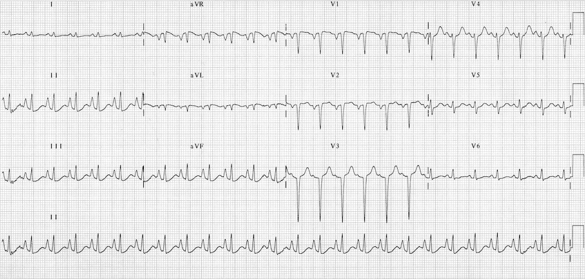
Dr. Smith's ECG Blog ECG with Aslanger's Pattern. CT Pulmonary
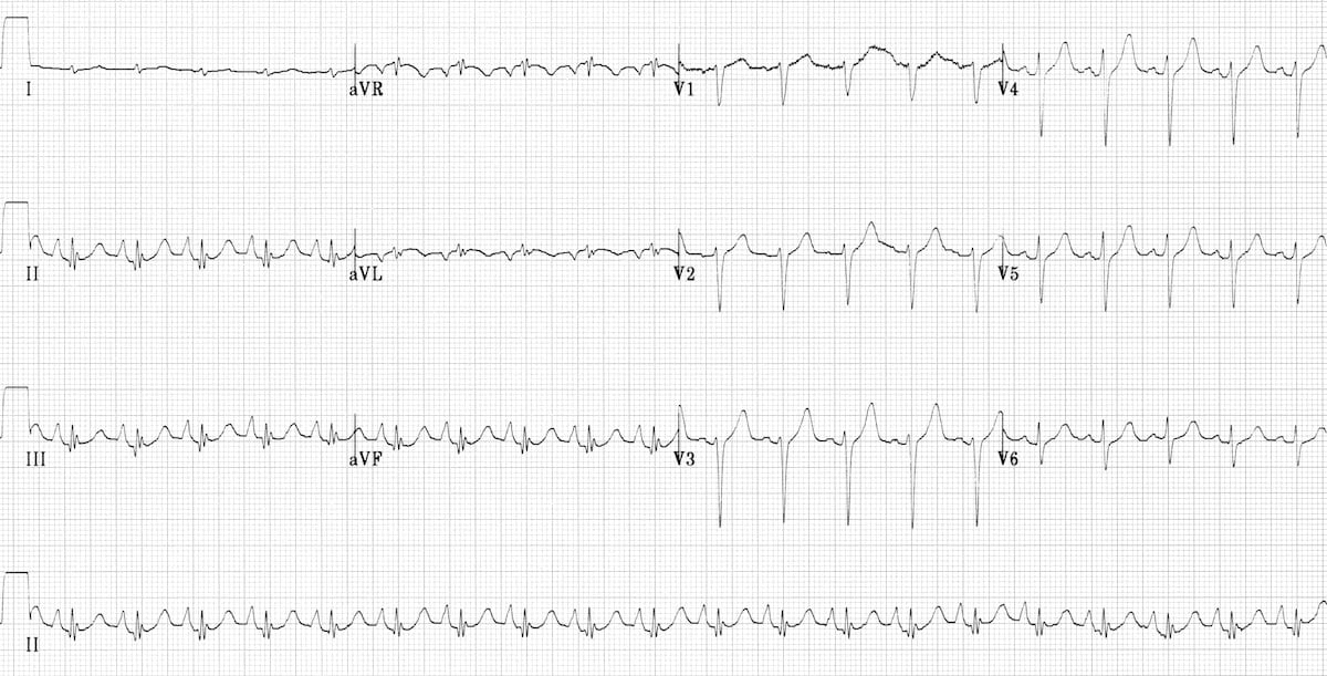
Longembolie Ecg / Pulmonary Pressures and ECG Patterns EMS 12 Lead
pulmonary disease pattern ecg Hình ảnh có liên quan Diseases Club
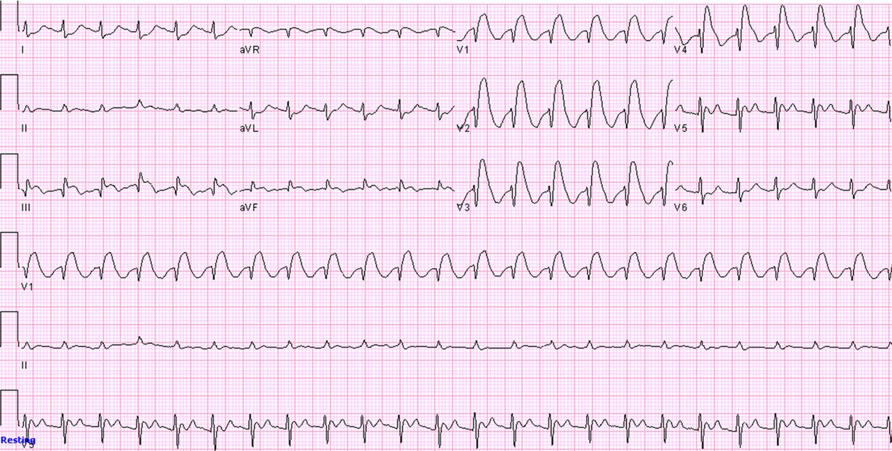
ECG in Chronic Obstructive Pulmonary Disease • LITFL • ECG Library

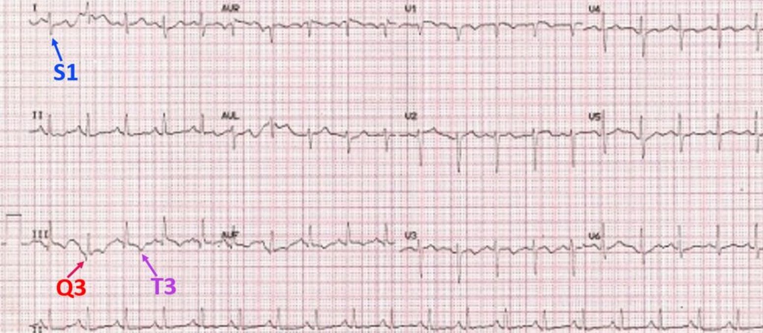
The ECG's of Pulmonary Embolism Resus
pulmonary disease pattern ecg Hình ảnh có liên quan Diseases Club
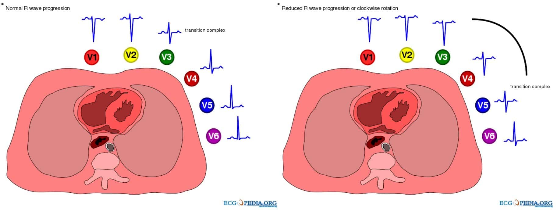
ECG in Chronic Obstructive Pulmonary Disease • LITFL • ECG Library
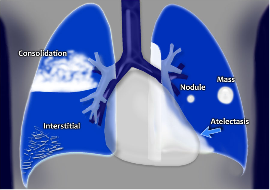
Chest XRay Lung disease FourPattern Approach NCLEX Quiz

Longembolie Ecg / Pulmonary Pressures and ECG Patterns EMS 12 Lead

Figure 2. Right bundle branch block (RBBB) and left bundle branch block
Ecg Findings Often Suggest Right Ventricular Pressure Overload Or Strain.
Web In Copd, The Various Pathophysiological Mechanisms Modify The Ecg Differently.
Our Aim Was To Separate The Effects On Ecg By Airway Obstruction, Emphysema And Right Ventricular (Rv) Afterload In Patients With Copd.
Related Post: